
Incisions
I close almost all of my incisions with sutures that are under the skin, just like a movie star's face. This is called a "subcuticular" closure, which is just Latin for "under the skin" closure. Although this takes more time and effort (and most surgeons who operate on the hand do not do them, not even hand surgeons), I do it because I think it has several advantages. There are no sutures to be removed, the sutures are not visible and therefore do not frighten the patients, the incision heals better, and I think that there is less chance for infection.
Here is what your incision might look like after surgery:
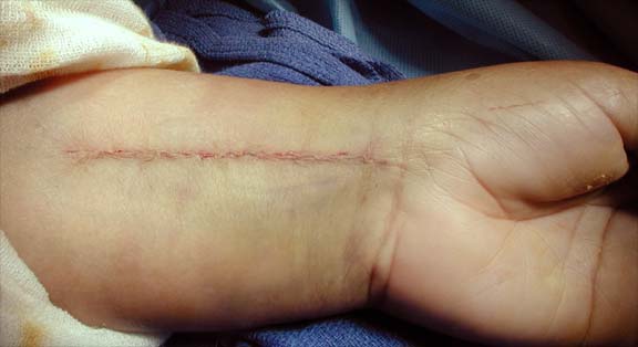
This is a picture, in the operating room, right after the skin is closed with a subcuticular closure and before the dressing is put on. Note that all the sutures are under the skin and you can't see them.
(If you want to see before and after pictures, click here, but it is rather graphic. I warned you.)
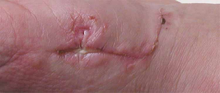
Now, let us look at some other incisions. This first one is a patient who is doing fairly well 10 days after carpal tunnel surgery, but he went back to work fixing diesel engines right away.
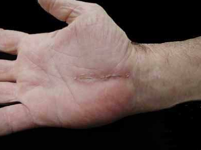
This is an incision for a carpal tunnel surgery at 10 days. For a closer look and a description of the incision, click here.
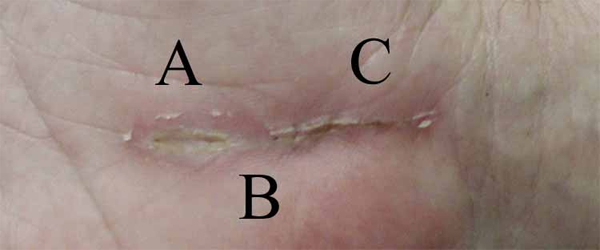
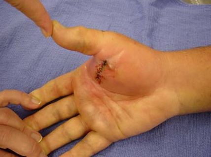
This is an incision for a thumb joint replacement surgery (LRTI). For a closer look and a description of the incision, click here.
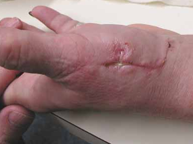
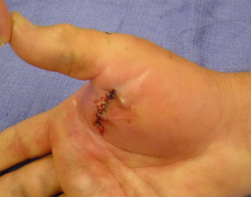
Here is an incision that is infected. The incision was made by an urgent care doctor. Click here to get more history and a closer view.
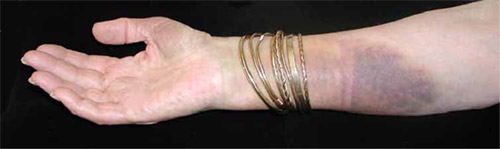
Incisions will usually develop some bruising. The technical name for this is "ecchymosis"
Luckily, infected incisions from hand surgery are very rare, and I have only had about eight in 21 years. They are so rare, in fact, that I don't have any pictures of one.
Visible Suture or White Fluid Around Sutures
Sometimes patients can see little bits of white suture sticking out of the wound. This is not a problem. It just means that the body is getting rid of the suture by pushing it out rather than absorbing it. Do not worry, just wash the incision with soap and the suture bits will wash off.
Sometimes patients can express (this means "push out") some white fluid that looks like pus. It is very unlikely that this is actually pus. If you are interested in what is going on in your incision, please read on!
White Blood Cells
There are at least six kinds of white blood cells.
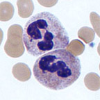 |
This is the most common kind of white cell, a neutrophil. Its job is to attack bacteria. Pus is composed of this kind of white blood cell. |
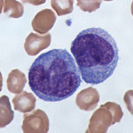 |
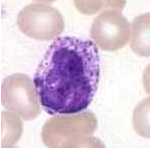
This white blood cell is a monocyte and is the precursor to a giant cell. |
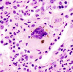 |
This is a giant cell, the kind of white blood cell that comes from monocytes and are the main fighters against foreign material in your body. Sutures are viewed by the body as "foreign material", and lots of giant celll can accumulate around the suture. They would be then termed "foreign body giant cells". |
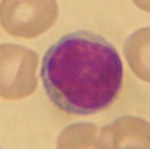 |
This white blood cell is a lympocyte, involved in producing antibodies. |
 |
This white blood cell is a basophil, involved in histamine release. When you take an antihistamine, you are trying to calm down these cells. |
| You can read more about white cells on Wikipedia. |
Pus is a collection of white cells of several kinds, just as illustrated above. Most of pus is made up of neutrophils, the first kind of white cell listed above. They are also called PMN's (which stands for polymorphonuclear leukocytes, so you can see why we call them PMN's!)
The white fluid that collects around the suture is actuallly a different kind of white fluid than pus. You can only tell by looking through a microscope, but the fluid around the sutures is composed of foreign-body giant cells. They look to the naked eye like PMN's and are closely related, but are just the way the body dissolves the suture. There is no infection, so the fluid around the sutures is called a "sterile stitch abscess." In some ways this is a bad name, since it is not really an abscess. The term, however, is too well established to be changed.
Sterile Stitch Abscess
Here is a great picture, from Wikipedia, of a sterile stitch abscess. The photograph was taken at lower power than the pictures above, to show you a larger area:
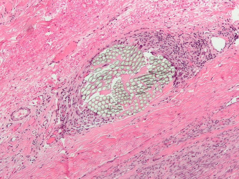
The green material is the suture (it has many strands, for strength and flexibility). The dark blue small cells are the foreign-body giant cells, which look like pus to the patient. The pink cells are muscle cells.
If you find that you can see the white sutures that I used to close your incision, don't worry. Your body will absorb the deeper parts of the suture, and you can clean your skin with soap and the suture bits will wash away.
You can tell if you are getting a real infection if the skin and surgical site are increasingly very tender, the skin becomes very red (not mildly pink, which is normal; look at the pictures above), and constantly draining fluid. If you have a question, call me and I can look at it. Remember, infections after my surgeries are very rare: I have had about 8 in 21 years.
| Would you like to search the medical library of the National Library of Medicine for scientific papers on this topic? Just click on the Pub - Med image: | |
| Remember the admonition from the Patient Education Links Page: the Internet has a lot of information, much of it incorrect. I have reviewed the sites that I have linked to, and have only linked to sites when I personally know the surgeon who posted it, or am a member of the organization that posted it. However, I may not agree with all that is on that site, and it may have changed since I reviewed it. If any of the information is not consistent with what I have told you, please download the material and bring it in. | |